CONTENTS
Introduction
The South Atlantic Bight
Methods
Octocoral Morphology
Glossary
Gorgonacean
Bauplan
see this for keys
Notes on the Species
Carijoa
riisei
Scleranthelia
rugosa
Telesto fruticulosa
Telesto nelleae
Telesto sanguinea
Bellonella rubistella
Pseudodrifa nigra
Nidalia occidentalis
Iciligorgia schrammi
Diodogorgia
nodulifera
Titanideum
frauenfeldii
Muricea pendula
Thesea nivea
Bebryce cinerea
Bebryce parastellata
Scleracis guadalupensis
Paramuricea sp.
Leptogorgia hebes
Leptogorgia punicea
Leptogorgia
cardinalis
Leptogorgia virgulata
Leptogorgia setacea
Leptogorgia euryale
Viminella
barbadensis
Renilla reniformis
Sclerobelemnon
theseus
Stylatula elegans
Virgularia presbytes
| Guide
to the Shallow Water (0-200 m) Octocorals of the South Atlantic
Bight.
S. T. DeVictor
& S. L. Morton, 2007
Telesto sanguinea
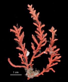
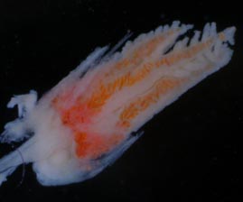
Deichmann, 1936 Remarks. Telesto sanguinea colonies are monopodially branched and may have multiple branches rising from stolons. The daughter polyps sometimes develop into tertiary branches. The color of the coenenchyme is bright red but may be obscured or completely encrusted by fouling organisms such as sponges and bryozoans. The species may rarely be orange, pink or yellow (Bayer 1961). As is typical of the members of this genus in the Atlantic, there are eight longitudinal grooves present in the body wall of the primary polyp but they are sometimes more distinct near the calyces or the base of the colony. Atlantic distribution: South Carolina to the Florida
Keys and Gulf of Mexico, 18-134 m (Deichmann, 1936; Bayer, 1961;
NMNH collections; SERTC collection). |
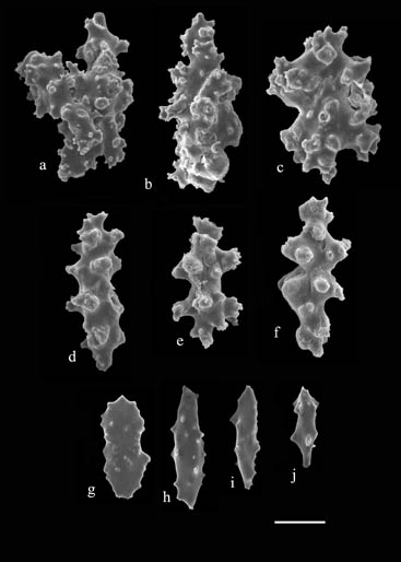
Figure 3. Sclerites of Telesto sanguinea (USNM 50357). a) fused sclerites from body wall; b) sclerite from body wall; c-f) sclerites from calyx wall; g-j) rods from polyp (scale bar = 50 µm). |
 |


