CONTENTS
Introduction
The South Atlantic Bight
Methods
Octocoral Morphology
Glossary
Gorgonacean
Bauplan
see this for keys
Notes on the Species
Carijoa
riisei
Scleranthelia
rugosa
Telesto fruticulosa
Telesto nelleae
Telesto sanguinea
Bellonella rubistella
Pseudodrifa nigra
Nidalia occidentalis
Iciligorgia schrammi
Diodogorgia
nodulifera
Titanideum
frauenfeldii
Muricea pendula
Thesea nivea
Bebryce cinerea
Bebryce parastellata
Scleracis guadalupensis
Paramuricea sp.
Leptogorgia hebes
Leptogorgia punicea
Leptogorgia
cardinalis
Leptogorgia virgulata
Leptogorgia setacea
Leptogorgia euryale
Viminella
barbadensis
Renilla reniformis
Sclerobelemnon
theseus
Stylatula elegans
Virgularia presbytes
| Guide
to the Shallow Water (0-200 m) Octocorals of the South Atlantic
Bight.
S. T. DeVictor
& S. L. Morton, 2007
Telesto fruticulosa
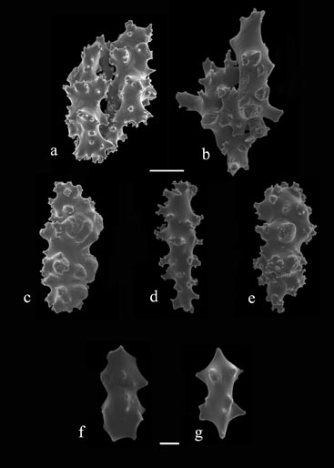
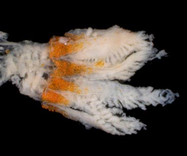
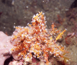
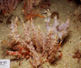
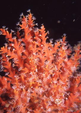
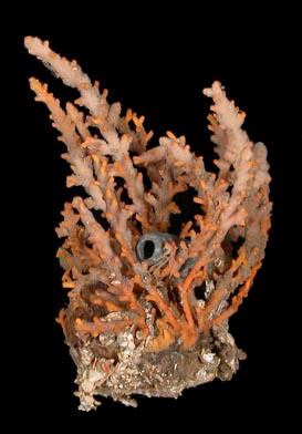
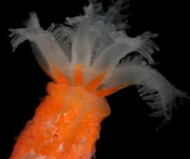
Dana, 1846 Remarks. Telesto fruticulosa colonies are monopodially branched and usually found in colonies of multiple branches. The daughter polyps sometimes develop into tertiary branches. The color of the coenenchyme may be orange, light red, or yellow, but may be obscured or completely encrusted by fouling organisms such as sponges and bryozoans. One encrusting sponge produced a thin veneer that was observed to be the bright red of Telesto sanguinea, a species closely resembling T. fruticulosa, such that the true color of the colony was completely obscured until preserved in ethanol. As is typical of the members of this genus in the Atlantic, there are eight longitudinal grooves present in the body wall of the primary polyps, but they are sometimes more distinct near the calyces or the base of the colony. There is a dense cluster of vertically oriented, overlapping flat rods in the base and proximal half of the polyp tentacles. Atlantic distribution: Coasts of the North Carolina,
South Carolina, Georgia and northern Florida, 7-100 m (Deichmann,
1936; Bayer 1961; NMNH collections; SERTC collection).
|
|
 |
